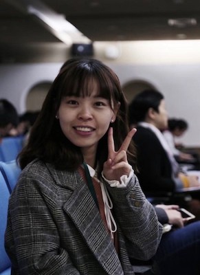
金璐红
一 教师基本信息
姓名:金璐红
学历/职称:博士 副教授
邮箱:luhongjin@djbet88.net
指导专业:计算机科学与技术,电子信息
二 研究领域及方向
显微成像技术研究及图像定量分析。在学习了解现有显微成像技术及其局限性的基础上,设计图像处理算法实现成像技术的优化,并为后续开展相关的细胞生物学研究提供技术支持。
三 教育及工作经历
201009—201406 中国计量大学 信息科学学院 学士
201509—202009 浙江大学 生物医学工程与仪器科学学院 博士
201811—201910 美国北卡罗来纳大学教堂山分校 药理系 访问学者
202009—202310 浙江大学 生物医学工程与仪器科学学院 博士后
202311—至今 杭州师范大学 电子竞技博彩 副教授
四 学术简介
从事显微成像技术研究及图像定量分析。主要针对现有的传统超分辨成像技术存在原始数据需求量大、荧光漂白严重、成像延时以及荧光标记种类有限等问题,利用深度学习技术设计相应的图像处理算法实现成像技术的优化。与此同时,也关注于利用现有成像技术的优势,结合显微亚细胞结构各自的特点,设计对应的亚细胞结构三维重建算法以及单粒子追踪算法,为后续开展相关的细胞生物学研究提供技术支持。
作为项目负责人主持国家自然科学青年基金项目1项,中国博士后面上基金项目1项,浙江省自然科学基金探索项目1项。曾参与科技部973项目、国家重点研发计划与基金委重大仪器专项等多项国家和省部级纵向研究课题。共参与发表了25篇研究论文,其中以第一作者/共一及共通讯身份发表于《Nature Communications》、《Optics and Lasers in Engineering》、《Computers in Biology and Medicine》等业内权威期刊8篇。曾2次在相关国际学术会议上做口头报告,申请并获批4项国家发明专利、1项实用新型专利以及3项软件著作权。
五 科研成果
【科研项目】
[1] 基于深度学习的快速结构光照明超分辨显微成像技术研究,国家自然科学基金委员会, 青年科学基金项目, 62105288, 2022-01-01至2024-12-31,主持
[2] 基于深度学习的智能超分辨显微成像系统研究,浙江省自然科学基金,探索项目, LQ22F050018, 2022-01-01至2024-12-31,主持
[3] 深度学习驱动的多目标快速结构光照明显微成像辅助系统的研究,中国博士后面上基金项目,2021M692831,2021-07-01至2023-06-30,主持
【发表论文】
[1] Ye Z, Huang Y, Zhang J, Chen Y, Ye H, Ji C, Jin L, Gan Y, Sun Y, Tao W, Han Y, Liu X, Chen Y*, Kuang C*, Liu W*. Universal and High-Fidelity Resolution Extending for Fluorescence Microscopy Using a Single-Training Physics-Informed Sparse Neural Network. Intelligent Computing. 2024;3:0082.
[2] Chen J#, Fang Q#, Huang L, Xin Ye, Jin L*, Zhang H, Luo Y, Zhu M, Zhang L, Ji B, Tian X*, Xu Y*. Deep-Learning Accelerated Super-Resolution Radial Fluctuations Enables Real-time Live Cell Imaging. Optics and Lasers in Engineering. 2024, 172, 107840.
[3] Zhu M#, Zhang L#, Jin L, Chen Y, Yang H, Xu Y*. Fast DNA-PAINT Imaging of Cellular Structures with U-Net. Biophysics Reports. 2024, 9(4):1-11.
[4] Yang H, Yu J, Jin L, Zhao Y, Gao Q, Shi C, Ye L, Li D, Yu H and Xu Y*. A deep learning based method for automatic analysis of high-throughput droplet digital PCR images. Analyst. 2022, 148(2), 239:247.
[5] Yang H, Zhu Y, Yu J, Jin L, Guo Z, Zheng C, Fu J, Xu Y*. Boosting Microscopic Object Detection via Feature Activation Map Guided Poisson Blending. Mathematical Biosciences and Engineering. 2023, 20(10): 18301-18317.
[6] Xu Y, Jin L, Toomre D. Imaging Single-Vesicle Exocytosis with Total Internal Reflection Fluorescence Microscopy (TIRFM). Methods in Molecular Biology, 2022, 2473, 157-164.
[7] Liu Z, Zhang H, Jin L, Chen J, Nedzved A, Ablameyko S, Ma Q, Yu J, Xu Y*. U-Net-based deep learning for tracking and quantitative analysis of intracellular vesicles in time-lapse microscopy images. Journal of Innovative Optical Health Sciences. 2022, 15(5), 2250031.
[8] Zhu M#, Zhang L#, Jin L, Chen J, Zhang Y, Xu Y*. DNA-PAINT Imaging Accelerated by Machine Learning. Frontiers in Chemistry. 2022, 10, 864701.
[9] Li X, Xie S, Liu W, Jin L, Xu Y, Zhang L, Hao X, Han Y, Kuang. Speckle-free laser projection structured illumination microscopy based on a digital micromirror device. Optics Express, 2021, 29(26), 43917-43928.
[10] Liu Z, Jin J, Nedzved A, Ablameyko S, Xu Y*. Deep Learning for Tracking of Intracellular Vesicles in Time-lapse Microscopy Images. Proc. SPIE 12069, AOPC 2021.
[11] Liu Z#, Jin L#, Chen J, Fang Q, Ablameyko S, Yin Z, Xu Y*. A survey on applications of deep learning in microscopy image analysis. Computers in Biology and Medicine. 2021, 134, 104523.
[12] Jin L#, Liu B#,*, Zhao F, Hahn S, Dong B, Song R, Elston T, Xu Y*, Hahn K*. Deep learning enables structured illumination microscopy with low light levels and enhanced speed. Nature Communications. 2020, 11: 1934.
[13] Jin L, Zhao F, Lin W, Zhou X, Kuang C, Nedzved A, Ablameyko S, Liu X, Xu Y*. Development of Fan-shaped Tracker (FsT) for Single Particle Tracking. Microscopy Research and Technique. 2020.
[14] Jin L, Zhao F, Lin W, Zhou X, Kuang C, Xu L, Ablameyko S, Xu Y*. Fan-shaped tracker (FsT) for particle trajectory reconstruction. Optics in Health Care and Biomedical Optics IX 2019. 2019, 111900Y.
[15] Nedzved O, Jin L, Nedzved A*, Lin W, Ablameyko S, Xu Y. Automatic analysis of moving particles by total internal reflection fluorescence microscopy. Pattern Recognition and Information Processing 2019, 1055(1):228-239.
[16] Chen Y, Liu W, Zhang Z, Zheng C, Huang Y, Cao R, Zhu D, Xu L, Meng Z, Zhang YH, Fan J, Jin L, Xu Y, Kuang C*, Liu X*. Multi-color live-cell super-resolution volume imaging with multi-angle interference microscopy. Nature Communications 2018, 9:4818.
[17] Zhou X, Wang J, Chen J, Qi Y, Nan D, Jin L, Qian X, Wang X, Chen Q*, Liu X, Xu Y*. Optogenetic control of epithelial-mesenchymal transition in cancer cells. Scientific Reports 2018, 8:14098.
[18] Zheng C, Zhao G, Liu W, Chen Y, Zhang Z, Jin L, Xu Y, Kuang C*, Liu X. Three-dimensional super-resolved live cell imaging through polarized multi-angle TIRF. Optics Letters 2018, 43:1423-1426.
[19] Jin L, Zhou X, Xiu P, Luo W, Huang Y, Yu F, Kuang C, Sun Y, Liu X, Xu Y*. Imaging and reconstruction of cell cortex structures near the cell surface. Optics Communications 2017, 402:699-705.
[20] Jin L, Wu J, Xiu P, Fan J, Hu M, Kuang C, Xu Y*, Zheng X, Liu X. High-resolution 3-D reconstruction of microtubule structures by quantitative multi-angle total internal reflection fluorescence microscopy. Optics Communications 2017, 395:16-23.
[21] Nedzved O, Jin L, Nedzved A*, Lin W, Ablameyko S, Xu Y*. Automatic Analysis of Moving Particles by Total Internal Reflection Fluorescence Microscopy. Communications in Computer and Information Science, 14th International conference on Pattern Recognition and Information Processing, PRIP 2019. 2019, 1055:228-239.
[22] 林婉妮,金璐红,许迎科*. 超分辨显微成像中荧光单分子定位算法中的研究进展. 中国生物医学工程学报. 2020,39(2):93-101.
[23] Jin L, Xiu P, Zhou X, Fan J, Kuang C, Liu X, Xu Y*. 3D reconstruction of cortical microtubules using multi-angle total internal reflection fluorescence microscopy. International Conference on Innovative Optical Health Science 2017, 10245:1024506.
[24] Huang Y, Zhu D, Jin L, Kuang C, Xu Y*, Liu X. Laser scanning saturated structured illumination microscopy based on phase modulation. Optics Communications 2017, 396:262-266.
[25] Wu J, Xiu P, Jin L, Nan D, Kuang C, Zheng X, Xu Y*, Liu X. A new method for axial decay function calibration of evanescent field in multi-angle total internal reflection fluorescence microscopy. Journal of Physics: Conference Series 2016, 680 (1):012025.
【专利】
[1] 许迎科,张恒,金璐红.一种荧光显微图像超分辨重建方法和装置以及计算设备, ZL202210558421.0,2023年9月8日,发明专利
[2] 许迎科,杨海旭,金璐红.一种微液滴数字PCR液滴检测方法及系统, ZL202210442041.0,2022年8月5日,发明专利
[3] 许迎科,金璐红,刘旭.基于深度学习的荧光显微图像超分辨重建技术, ZL202010166665.5,2022年4月29日,发明专利
[4] 许迎科,金璐红,刘旭.基于显微图像的亚细胞结构运动轨迹确定方法, ZL201811268090.7,2020年9月1日,发明专利
[5] 许迎科,金璐红,樊剑南,南迪,孙永红,匡翠方,刘旭. 新型多角度环状光学照明显微成像系统,ZL201720543615.8,2017年12月15日,实用新型专利
[6] 许迎科,金璐红,刘旭. 全内反射荧光显微图像的三维重建及图像定量分析系统V1.0, 2017SR399591, 2017年7月26日,软件著作权
[7] 许迎科,刘智超,金璐红. 荧光显微图像中亚细胞结构运动模拟系统V1.0, 2021SR1011058,2021年3月10日,软件著作权
[8] 许迎科,刘智超,金璐红. 荧光显微图像中亚细胞结构识别与追踪系统V1.0, 2021SR0980582,2021年3月15日,软件著作权
欢迎积极、主动、严于律己的学生加入课题组。


